Sunday, 16 January 2011 16:18

Introduction and Concepts

Rate this item
(3 votes)

Mechanistic toxicology is the study of how chemical or physical agents interact with living organisms to cause toxicity. Knowledge of the mechanism of toxicity of a substance enhances the ability to prevent toxicity and design more desirable chemicals; it constitutes the basis for therapy upon overexposure, and frequently enables a further understanding of fundamental biological processes. For purposes of this Encyclopaedia the emphasis will be placed on animals to predict human toxicity. Different areas of toxicology include mechanistic, descriptive, regulatory, forensic and environmental toxicology (Klaassen, Amdur and Doull 1991). All of these benefit from understanding the fundamental mechanisms of toxicity.

Why Understand Mechanisms of Toxicity?

Understanding the mechanism by which a substance causes toxicity enhances different areas of toxicology in different ways. Mechanistic understanding helps the governmental regulator to establish legally binding safe limits for human exposure. It helps toxicologists in recommending courses of action regarding clean-up or remediation of contaminated sites and, along with physical and chemical properties of the substance or mixture, can be used to select the degree of protective equipment required. Mechanistic knowledge is also useful in forming the basis for therapy and the design of new drugs for treatment of human disease. For the forensic toxicologist the mechanism of toxicity often provides insight as to how a chemical or physical agent can cause death or incapacitation.

If the mechanism of toxicity is understood, descriptive toxicology becomes useful in predicting the toxic effects of related chemicals. It is important to understand, however, that a lack of mechanistic information does not deter health professionals from protecting human health. Prudent decisions based on animal studies and human experience are used to establish safe exposure levels. Traditionally, a margin of safety was established by using the “no adverse effect level” or a “lowest adverse effect level” from animal studies (using repeated-exposure designs) and dividing that level by a factor of 100 for occupational exposure or 1,000 for other human environmental exposure. The success of this process is evident from the few incidents of adverse health effects attributed to chemical exposure in workers where appropriate exposure limits had been set and adhered to in the past. In addition, the human lifespan continues to increase, as does the quality of life. Overall the use of toxicity data has led to effective regulatory and voluntary control. Detailed knowledge of toxic mechanisms will enhance the predictability of newer risk models currently being developed and will result in continuous improvement.

Understanding environmental mechanisms is complex and presumes a knowledge of ecosystem disruption and homeostasis (balance). While not discussed in this article, an enhanced understanding of toxic mechanisms and their ultimate consequences in an ecosystem would help scientists to make prudent decisions regarding the handling of municipal and industrial waste material. Waste management is a growing area of research and will continue to be very important in the future.

Techniques for Studying Mechanisms of Toxicity

The majority of mechanistic studies start with a descriptive toxicological study in animals or clinical observations in humans. Ideally, animal studies include careful behavioural and clinical observations, careful biochemical examination of elements of the blood and urine for signs of adverse function of major biological systems in the body, and a post-mortem evaluation of all organ systems by microscopic examination to check for injury (see OECD test guidelines; EC directives on chemical evaluation; US EPA test rules; Japan chemicals regulations). This is analogous to a thorough human physical examination that would take place in a hospital over a two- to three-day time period except for the post-mortem examination.

Understanding mechanisms of toxicity is the art and science of observation, creativity in the selection of techniques to test various hypotheses, and innovative integration of signs and symptoms into a causal relationship. Mechanistic studies start with exposure, follow the time-related distribution and fate in the body (pharmacokinetics), and measure the resulting toxic effect at some level of the system and at some dose level. Different substances can act at different levels of the biological system in causing toxicity.

Exposure

The route of exposure in mechanistic studies is usually the same as for human exposure. Route is important because there can be effects that occur locally at the site of exposure in addition to systemic effects after the chemical has been absorbed into the blood and distributed throughout the body. A simple yet cogent example of a local effect would be irritation and eventual corrosion of the skin following application of strong acid or alkaline solutions designed for cleaning hard surfaces. Similarly, irritation and cellular death can occur in cells lining the nose and/or lungs following exposure to irritant vapours or gases such as oxides of nitrogen or ozone. (Both are constituents of air pollution, or smog). Following absorption of a chemical into blood through the skin, lungs or gastrointestinal tract, the concentration in any organ or tissue is controlled by many factors which determine the pharmacokinetics of the chemical in the body. The body has the ability to activate as well as detoxify various chemicals as noted below.

Role of Pharmacokinetics in Toxicity

Pharmacokinetics describes the time relationships for chemical absorption, distribution, metabolism (biochemical alterations in the body) and elimination or excretion from the body. Relative to mechanisms of toxicity, these pharmacokinetic variables can be very important and in some instances determine whether toxicity will or will not occur. For instance, if a material is not absorbed in a sufficient amount, systemic toxicity (inside the body) will not occur. Conversely, a highly reactive chemical that is detoxified quickly (seconds or minutes) by digestive or liver enzymes may not have the time to cause toxicity. Some polycyclic halogenated substances and mixtures as well as certain metals like lead would not cause significant toxicity if excretion were rapid; but accumulation to sufficiently high levels determines their toxicity since excretion is not rapid (sometimes measured in years). Fortunately, most chemicals do not have such long retention in the body. Accumulation of an innocuous material still would not induce toxicity. The rate of elimination from the body and detoxication is frequently referred to as the half-life of the chemical, which is the time for 50% of the chemical to be excreted or altered to a non-toxic form.

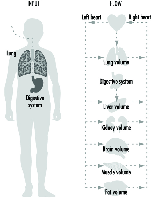
However, if a chemical accumulates in a particular cell or organ, that may signal a reason to further examine its potential toxicity in that organ. More recently, mathematical models have been developed to extrapolate pharmacokinetic variables from animals to humans. These pharmacokinetic models are extremely useful in generating hypotheses and testing whether the experimental animal may be a good representation for humans. Numerous chapters and texts have been written on this subject (Gehring et al. 1976; Reitz et al. 1987; Nolan et al. 1995). A simplified example of a physiological model is depicted in figure 1.

Figure 1. A simplified pharmacokinetic model

TOX210F1

Different Levels and Systems Can Be Adversely Affected

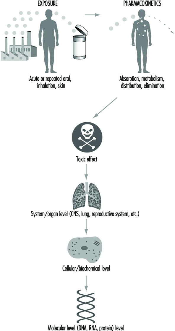
Toxicity can be described at different biological levels. Injury can be evaluated in the whole person (or animal), the organ system, the cell or the molecule. Organ systems include the immune, respiratory, cardiovascular, renal, endocrine, digestive, muscolo-skeletal, blood, reproductive and central nervous systems. Some key organs include the liver, kidney, lung, brain, skin, eyes, heart, testes or ovaries, and other major organs. At the cellular/biochemical level, adverse effects include interference with normal protein function, endocrine receptor function, metabolic energy inhibition, or xenobiotic (foreign substance) enzyme inhibition or induction. Adverse effects at the molecular level include alteration of the normal function of DNA-RNA transcription, of specific cytoplasmic and nuclear receptor binding, and of genes or gene products. Ultimately, dysfunction in a major organ system is likely caused by a molecular alteration in a particular target cell within that organ. However, it is not always possible to trace a mechanism back to a molecular origin of causation, nor is it necessary. Intervention and therapy can be designed without a complete understanding of the molecular target. However, knowledge about the specific mechanism of toxicity increases the predictive value and accuracy of extrapolation to other chemicals. Figure 2 is a diagrammatic representation of the various levels where interference of normal physiological processes can be detected. The arrows indicate that the consequences to an individual can be determined from top down (exposure, pharmaco- kinetics to system/organ toxicity) or from bottom up (molecular change, cellular/biochemical effect to system/organ toxicity).

Figure 2. Reresentation of mechanisms of toxicity

TOX210F2

Examples of Mechanisms of Toxicity

Mechanisms of toxicity can be straightforward or very complex. Frequently, there is a difference among the type of toxicity, the mechanism of toxicity, and the level of effect, related to whether the adverse effects are due to a single, acute high dose (like an accidental poisoning), or a lower-dose repeated exposure (from occupational or environmental exposure). Classically, for testing purposes, an acute, single high dose is given by direct intubation into the stomach of a rodent or exposure to an atmosphere of a gas or vapour for two to four hours, whichever best resembles the human exposure. The animals are observed over a two-week period following exposure and then the major external and internal organs are examined for injury. Repeated-dose testing ranges from months to years. For rodent species, two years is considered a chronic (lifetime) study sufficient to evaluate toxicity and carcinogenicity, whereas for non-human primates, two years would be considered a subchronic (less than lifetime) study to evaluate repeated dose toxicity. Following exposure a complete examination of all tissues, organs and fluids is conducted to determine any adverse effects.

Acute Toxicity Mechanisms

The following examples are specific to high-dose, acute effects which can lead to death or severe incapacitation. However, in some cases, intervention will result in transient and fully reversible effects. The dose or severity of exposure will determine the result.

Simple asphyxiants. The mechanism of toxicity for inert gases and some other non-reactive substances is lack of oxygen (anoxia). These chemicals, which cause deprivation of oxygen to the central nervous system (CNS), are termed simple asphyxiants. If a person enters a closed space that contains nitrogen without sufficient oxygen, immediate oxygen depletion occurs in the brain and leads to unconsciousness and eventual death if the person is not rapidly removed. In extreme cases (near zero oxygen) unconsciousness can occur in a few seconds. Rescue depends on rapid removal to an oxygenated environment. Survival with irreversible brain damage can occur from delayed rescue, due to the death of neurons, which cannot regenerate.

Chemical asphyxiants. Carbon monoxide (CO) competes with oxygen for binding to haemoglobin (in red blood cells) and therefore deprives tissues of oxygen for energy metabolism; cellular death can result. Intervention includes removal from the source of CO and treatment with oxygen. The direct use of oxygen is based on the toxic action of CO. Another potent chemical asphyxiant is cyanide. The cyanide ion interferes with cellular metabolism and utilization of oxygen for energy. Treatment with sodium nitrite causes a change in haemoglobin in red blood cells to methaemoglobin. Methaemoglobin has a greater binding affinity to the cyanide ion than does the cellular target of cyanide. Consequently, the methaemoglobin binds the cyanide and keeps the cyanide away from the target cells. This forms the basis for antidotal therapy.

Central nervous system (CNS) depressants. Acute toxicity is characterized by sedation or unconsciousness for a number of materials like solvents which are not reactive or which are transformed to reactive intermediates. It is hypothesized that sedation/anaesthesia is due to an interaction of the solvent with the membranes of cells in the CNS, which impairs their ability to transmit electrical and chemical signals. While sedation may seem a mild form of toxicity and was the basis for development of the early anaesthetics, “the dose still makes the poison”. If sufficient dose is administered by ingestion or inhalation the animal can die due to respiratory arrest. If anaesthetic death does not occur, this type of toxicity is usually readily reversible when the subject is removed from the environment or the chemical is redistributed or eliminated from the body.

Skin effects. Adverse effects to the skin can range from irritation to corrosion, depending on the substance encountered. Strong acids and alkaline solutions are incompatible with living tissue and are corrosive, causing chemical burns and possible scarring. Scarring is due to death of the dermal, deep skin cells responsible for regeneration. Lower concentrations may just cause irritation of the first layer of skin.

Another specific toxic mechanism of skin is that of chemical sensitization. As an example, sensitization occurs when 2,4-dinitrochlorobenzene binds with natural proteins in the skin and the immune system recognizes the altered protein-bound complex as a foreign material. In responding to this foreign material, the immune system activates special cells to eliminate the foreign substance by release of mediators (cytokines) which cause a rash or dermatitis (see “Immunotoxicology”). This is the same reaction of the immune system when exposure to poison ivy occurs. Immune sensitization is very specific to the particular chemical and takes at least two exposures before a response is elicited. The first exposure sensitizes (sets up the cells to recognize the chemical), and subsequent exposures trigger the immune system response. Removal from contact and symptomatic therapy with steroid-containing anti-inflammatory creams are usually effective in treating sensitized individuals. In serious or refractory cases a systemic acting immunosuppresant like prednisone is used in conjunction with topical treatment.

Lung sensitization. An immune sensitization response is elicited by toluene diisocyanate (TDI), but the target site is the lungs. TDI over-exposure in susceptible individuals causes lung oedema (fluid build-up), bronchial constriction and impaired breathing. This is a serious condition and requires removing the individual from potential subsequent exposures. Treatment is primarily symptomatic. Skin and lung sensitization follow a dose response. Exceeding the level set for occupational exposure can cause adverse effects.

Eye effects. Injury to the eye ranges from reddening of the outer layer (swimming-pool redness) to cataract formation of the cornea to damage to the iris (coloured part of the eye). Eye irritation tests are conducted when it is believed serious injury will not occur. Many of the mechanisms causing skin corrosion can also cause injury to the eyes. Materials corrosive to the skin, like strong acids (pH less than 2) and alkali (pH greater than 11.5), are not tested in the eyes of animals because most will cause corrosion and blindness due to a mechanism similar to that which causes skin corrosion. In addition, surface active agents like detergents and surfactants can cause eye injury ranging from irritation to corrosion. A group of materials that requires caution is the positively charged (cationic) surfactants, which can cause burns, permanent opacity of the cornea and vascularization (formation of blood vessels). Another chemical, dinitrophenol, has a specific effect of cataract formation. This appears to be related to concentration of this chemical in the eye, which is an example of pharmacokinetic distributional specificity.

While the listing above is far from exhaustive, it is designed to give the reader an appreciation for various acute toxicity mechanisms.

Subchronic and Chronic Toxicity Mechanisms

When given as a single high dose, some chemicals do not have the same mechanism of toxicity as when given repeatedly as a lower but still toxic dose. When a single high dose is given, there is always the possibility of exceeding the person’s ability to detoxify or excrete the chemical, and this can lead to a different toxic response than when lower repetitive doses are given. Alcohol is a good example. High doses of alcohol lead to primary central nervous system effects, while lower repetitive doses result in liver injury.

Anticholinesterase inhibition. Most organophosphate pesticides, for example, have little mammalian toxicity until they are metabolically activated, primarily in the liver. The primary mechanism of action of organophosphates is the inhibition of acetylcholinesterase (AChE) in the brain and peripheral nervous system. AChE is the normal enzyme that terminates the stimulation of the neurotransmitter acetylcholine. Slight inhibition of AChE over an extended period has not been associated with adverse effects. At high levels of exposure, inability to terminate this neuronal stimulation results in overstimulation of the cholinergic nervous system. Cholinergic overstimulation ultimately results in a host of symptoms, including respiratory arrest, followed by death if not treated. The primary treatment is the administration of atropine, which blocks the effects of acetylcholine, and the administration of pralidoxime chloride, which reactivates the inhibited AChE. Therefore, both the cause and the treatment of organophosphate toxicity are addressed by understanding the biochemical basis of toxicity.

Metabolic activation. Many chemicals, including carbon tetrachloride, chloroform, acetylaminofluorene, nitrosamines, and paraquat are metabolically activated to free radicals or other reactive intermediates which inhibit and interfere with normal cellular function. At high levels of exposure this results in cell death (see “Cellular injury and cellular death”). While the specific interactions and cellular targets remain unknown, the organ systems which have the capability to activate these chemicals, like the liver, kidney and lung, are all potential targets for injury. Specifically, particular cells within an organ have a greater or lesser capacity to activate or detoxify these intermediates, and this capacity determines the intracellular susceptibility within an organ. Metabolism is one reason why an understanding of pharmacokinetics, which describes these types of transformations and the distribution and elimination of these intermediates, is important in recognizing the mechanism of action of these chemicals.

Cancer mechanisms. Cancer is a multiplicity of diseases, and while the understanding of certain types of cancer is increasing rapidly due to the many molecular biological techniques that have been developed since 1980, there is still much to learn. However, it is clear that cancer development is a multi-stage process, and critical genes are key to different types of cancer. Alterations in DNA (somatic mutations) in a number of these critical genes can cause increased susceptibility or cancerous lesions (see “Genetic toxic- ology”). Exposure to natural chemicals (in cooked foods like beef and fish) or synthetic chemicals (like benzidine, used as a dye) or physical agents (ultraviolet light from the sun, radon from soil, gamma radiation from medical procedures or industrial activity) are all contributors to somatic gene mutations. However, there are natural and synthetic substances (such as anti-oxidants) and DNA repair processes which are protective and maintain homeostasis. It is clear that genetics is an important factor in cancer, since genetic disease syndromes such as xeroderma pigmentosum, where there is a lack of normal DNA repair, dramatically increase susceptibility to skin cancer from exposure to ultraviolet light from the sun.

Reproductive mechanisms. Similar to cancer, many mechanisms of reproductive and/or developmental toxicity are known, but much is to be learned. It is known that certain viruses (such as rubella), bacterial infections and drugs (such as thalidomide and vitamin A) will adversely affect development. Recently, work by Khera (1991), reviewed by Carney (1994), show good evidence that the abnormal developmental effects in animal tests with ethylene glycol are attributable to maternal metabolic acidic metabolites. This occurs when ethylene glycol is metabolized to acid metabolites including glycolic and oxalic acid. The subsequent effects on the placenta and foetus appear to be due to this metabolic toxication process.

Conclusion

The intent of this article is to give a perspective on several known mechanisms of toxicity and the need for future study. It is important to understand that mechanistic knowledge is not absolutely necessary to protect human or environmental health. This knowledge will enhance the professional’s ability to better predict and manage toxicity. The actual techniques used in elucidating any particular mechanism depend upon the collective knowledge of the scientists and the thinking of those who make decisions regarding human health.


Back

Read 10863 times Last modified on Tuesday, 26 July 2022 19:33
More in this category: Cellular Injury and Cellular Death »

" DISCLAIMER: The ILO does not take responsibility for content presented on this web portal that is presented in any language other than English, which is the language used for the initial production and peer-review of original content. Certain statistics have not been updated since the production of the 4th edition of the Encyclopaedia (1998)."

Contents

Toxicology References

Andersen, KE and HI Maibach. 1985. Contact allergy predictive tests on guinea pigs. Chap. 14 in Current Problems in Dermatology. Basel: Karger.

Ashby, J and RW Tennant. 1991. Definitive relationships among chemical structure, carcinogenicity and mutagenicity for 301 chemicals tested by the US NTP. Mutat Res 257:229-306.

Barlow, S and F Sullivan. 1982. Reproductive Hazards of Industrial Chemicals. London: Academic Press.

Barrett, JC. 1993a. Mechanisms of action of known human carcinogens. In Mechanisms of Carcinogenesis in Risk Identification, edited by H Vainio, PN Magee, DB McGregor, and AJ McMichael. Lyon: International Agency for Research on Cancer (IARC).

—. 1993b. Mechanisms of multistep carcinogenesis and carcinogen risk assessment. Environ Health Persp 100:9-20.

Bernstein, ME. 1984. Agents affecting the male reproductive system: Effects of structure on activity. Drug Metab Rev 15:941-996.

Beutler, E. 1992. The molecular biology of G6PD variants and other red cell defects. Annu Rev Med 43:47-59.

Bloom, AD. 1981. Guidelines for Reproductive Studies in Exposed Human Populations. White Plains, New York: March of Dimes Foundation.

Borghoff, S, B Short and J Swenberg. 1990. Biochemical mechanisms and pathobiology of a-2-globulin nephropathy. Annu Rev Pharmacol Toxicol 30:349.

Burchell, B, DW Nebert, DR Nelson, KW Bock, T Iyanagi, PLM Jansen, D Lancet, GJ Mulder, JR Chowdhury, G Siest, TR Tephly, and PI Mackenzie. 1991. The UPD-glucuronosyltransferase gene superfamily: Suggested nomenclature based on evolutionary divergence. DNA Cell Biol 10:487-494.

Burleson, G, A Munson, and J Dean. 1995. Modern Methods in Immunotoxicology. New York: Wiley.

Capecchi, M. 1994. Targeted gene replacement. Sci Am 270:52-59.

Carney, EW. 1994. An integrated perspective on the developmental toxicity of ethylene glycol. Rep Toxicol 8:99-113.

Dean, JH, MI Luster, AE Munson, and I Kimber. 1994. Immunotoxicology and Immunopharmacology. New York: Raven Press.

Descotes, J. 1986. Immunotoxicology of Drugs and Chemicals. Amsterdam: Elsevier.

Devary, Y, C Rosette, JA DiDonato, and M Karin. 1993. NFkB activation by ultraviolet light not dependent on a nuclear signal. Science 261:1442-1445.

Dixon, RL. 1985. Reproductive Toxicology. New York: Raven Press.

Duffus, JH. 1993. Glossary for chemists of terms used in toxicology. Pure Appl Chem 65:2003-2122.

Elsenhans, B, K Schuemann, and W Forth. 1991. Toxic metals: Interactions with essential metals. In Nutrition, Toxicity and Cancer, edited by IR Rowland. Boca-Raton: CRC Press.

Environmental Protection Agency (EPA). 1992. Guidelines for exposure assessment. Federal Reg 57:22888-22938.

—. 1993. Principles of neurotoxicity risk assessment. Federal Reg 58:41556-41598.

—. 1994. Guidelines for Reproductive Toxicity Assessment. Washington, DC: US EPA: Office of Research and Development.

Fergusson, JE. 1990. The Heavy Elements. Chap. 15 in Chemistry, Environmental Impact and Health Effects. Oxford: Pergamon.

Gehring, PJ, PG Watanabe, and GE Blau. 1976. Pharmacokinetic studies in evaluation of the toxicological and environmental hazard of chemicals. New Concepts Saf Eval 1(Part 1, Chapter 8):195-270.

Goldstein, JA and SMF de Morais. 1994. Biochemistry and molecular biology of the human CYP2C subfamily. Pharmacogenetics 4:285-299.

Gonzalez, FJ. 1992. Human cytochromes P450: Problems and prospects. Trends Pharmacol Sci 13:346-352.

Gonzalez, FJ, CL Crespi, and HV Gelboin. 1991. cDNA-expressed human cytochrome P450: A new age in molecular toxicology and human risk assessment. Mutat Res 247:113-127.

Gonzalez, FJ and DW Nebert. 1990. Evolution of the P450 gene superfamily: Animal-plant “warfare,” molecular drive, and human genetic differences in drug oxidation. Trends Genet 6:182-186.

Grant, DM. 1993. Molecular genetics of the N-acetyltransferases. Pharmacogenetics 3:45-50.

Gray, LE, J Ostby, R Sigmon, J Ferrel, R Linder, R Cooper, J Goldman, and J Laskey. 1988. The development of a protocol to assess reproductive effects of toxicants in the rat. Rep Toxicol 2:281-287.

Guengerich, FP. 1989. Polymorphism of cytochrome P450 in humans. Trends Pharmacol Sci 10:107-109.

—. 1993. Cytochrome P450 enzymes. Am Sci 81:440-447.

Hansch, C and A Leo. 1979. Substituent Constants for Correlation Analysis in Chemistry and Biology. New York: Wiley.

Hansch, C and L Zhang. 1993. Quantitative structure-activity relationships of cytochrome P450. Drug Metab Rev 25:1-48.

Hayes AW. 1988. Principles and Methods of Toxicology. 2nd ed. New York: Raven Press.

Heindell, JJ and RE Chapin. 1993. Methods in Toxicology: Male and Female Reproductive Toxicology. Vol. 1 and 2. San Diego, Calif.: Academic Press.

International Agency for Research on Cancer (IARC). 1992. Solar and ultraviolet radiation. Lyon: IARC.

—. 1993. Occupational Exposures of Hairdressers and Barbers and Personal Use of Hair Colourants: Some Hair Dyes, Cosmetic Colourants, Industrial Dyestuffs and Aromatic Amines. Lyon: IARC.

—. 1994a. Preamble. Lyon: IARC.

—. 1994b. Some Industrial Chemicals. Lyon: IARC.

International Commission on Radiological Protection (ICRP). 1965. Principles of Environmental Monitoring Related to the Handling of Radioactive Materials. Report of Committee IV of The International Commission On Radiological Protection. Oxford: Pergamon.

International Program on Chemical Safety (IPCS). 1991. Principles and Methods for the Assessment of Nephrotoxicity Associated With Exposure to Chemicals, EHC 119. Geneva: WHO.

—. 1996. Principles and Methods for Assessing Direct Immunotoxicity Associated With Exposure to Chemicals, EHC 180. Geneva: WHO.

Johanson, G and PH Naslund. 1988. Spreadsheet programming - a new approach in physiologically based modeling of solvent toxicokinetics. Toxicol Letters 41:115-127.

Johnson, BL. 1978. Prevention of Neurotoxic Illness in Working Populations. New York: Wiley.

Jones, JC, JM Ward, U Mohr, and RD Hunt. 1990. Hemopoietic System, ILSI Monograph, Berlin: Springer Verlag.

Kalow, W. 1962. Pharmocogenetics: Heredity and the Response to Drugs. Philadelphia: WB Saunders.

—. 1992. Pharmocogenetics of Drug Metabolism. New York: Pergamon.

Kammüller, ME, N Bloksma, and W Seinen. 1989. Autoimmunity and Toxicology. Immune Dysregulation Induced By Drugs and Chemicals. Amsterdam: Elsevier Sciences.

Kawajiri, K, J Watanabe, and SI Hayashi. 1994. Genetic polymorphism of P450 and human cancer. In Cytochrome P450: Biochemistry, Biophysics and Molecular Biology, edited by MC Lechner. Paris: John Libbey Eurotext.

Kehrer, JP. 1993. Free radicals as mediators of tissue injury and disease. Crit Rev Toxicol 23:21-48.

Kellerman, G, CR Shaw, and M Luyten-Kellerman. 1973. Aryl hydrocarbon hydroxylase inducibility and bronochogenic carcinoma. New Engl J Med 289:934-937.

Khera, KS. 1991. Chemically induced alterations maternal homeostasis and histology of conceptus: Their etiologic significance in rat fetal anomalies. Teratology 44:259-297.

Kimmel, CA, GL Kimmel, and V Frankos. 1986. Interagency Regulatory Liaison Group workshop on reproductive toxicity risk assessment. Environ Health Persp 66:193-221.

Klaassen, CD, MO Amdur and J Doull (eds.). 1991. Casarett and Doull´s Toxicology. New York: Pergamon Press.

Kramer, HJ, EJHM Jansen, MJ Zeilmaker, HJ van Kranen and ED Kroese. 1995. Quantitative methods in toxicology for human dose-response assessment. RIVM-report nr. 659101004.

Kress, S, C Sutter, PT Strickland, H Mukhtar, J Schweizer, and M Schwarz. 1992. Carcinogen-specific mutational pattern in the p53 gene in ultraviolet B radiation-induced squamous cell carcinomas of mouse skin. Cancer Res 52:6400-6403.

Krewski, D, D Gaylor, M Szyazkowicz. 1991. A model-free approach to low-dose extrapolation. Env H Pers 90:270-285.

Lawton, MP, T Cresteil, AA Elfarra, E Hodgson, J Ozols, RM Philpot, AE Rettie, DE Williams, JR Cashman, CT Dolphin, RN Hines, T Kimura, IR Phillips, LL Poulsen, EA Shephare, and DM Ziegler. 1994. A nomenclature for the mammalian flavin-containing monooxygenase gene family based on amino acid sequence identities. Arch Biochem Biophys 308:254-257.

Lewalter, J and U Korallus. 1985. Blood protein conjugates and acetylation of aromatic amines. New findings on biological monitoring. Int Arch Occup Environ Health 56:179-196.

Majno, G and I Joris. 1995. Apoptosis, oncosis, and necrosis: An overview of cell death. Am J Pathol 146:3-15.

Mattison, DR and PJ Thomford. 1989. The mechanism of action of reproductive toxicants. Toxicol Pathol 17:364-376.

Meyer, UA. 1994. Polymorphisms of cytochrome P450 CYP2D6 as a risk factor in carcinogenesis. In Cytochrome P450: Biochemistry, Biophysics and Molecular Biology, edited by MC Lechner. Paris: John Libbey Eurotext.

Moller, H, H Vainio and E Heseltine. 1994. Quantitative estimation and prediction of risk at the International Agency for Research on Cancer. Cancer Res 54:3625-3627.

Moolenaar, RJ. 1994. Default assumptions in carcinogen risk assessment used by regulatory agencies. Regul Toxicol Pharmacol 20:135-141.

Moser, VC. 1990. Screening approaches to neurotoxicity: A functional observational battery. J Am Coll Toxicol 1:85-93.

National Research Council (NRC). 1983. Risk Assessment in the Federal Government: Managing the Process. Washington, DC: NAS Press.

—. 1989. Biological Markers in Reproductive Toxicity. Washington, DC: NAS Press.

—. 1992. Biologic Markers in Immunotoxicology. Subcommittee on Toxicology. Washington, DC: NAS Press.

Nebert, DW. 1988. Genes encoding drug-metabolizing enzymes: Possible role in human disease. In Phenotypic Variation in Populations, edited by AD Woodhead, MA Bender, and RC Leonard. New York: Plenum Publishing.

—. 1994. Drug-metabolizing enzymes in ligand-modulated transcription. Biochem Pharmacol 47:25-37.

Nebert, DW and WW Weber. 1990. Pharmacogenetics. In Principles of Drug Action. The Basis of Pharmacology, edited by WB Pratt and PW Taylor. New York: Churchill-Livingstone.

Nebert, DW and DR Nelson. 1991. P450 gene nomenclature based on evolution. In Methods of Enzymology. Cytochrome P450, edited by MR Waterman and EF Johnson. Orlando, Fla: Academic Press.

Nebert, DW and RA McKinnon. 1994. Cytochrome P450: Evolution and functional diversity. Prog Liv Dis 12:63-97.

Nebert, DW, M Adesnik, MJ Coon, RW Estabrook, FJ Gonzalez, FP Guengerich, IC Gunsalus, EF Johnson, B Kemper, W Levin, IR Phillips, R Sato, and MR Waterman. 1987. The P450 gene superfamily: Recommended nomenclature. DNA Cell Biol 6:1-11.

Nebert, DW, DR Nelson, MJ Coon, RW Estabrook, R Feyereisen, Y Fujii-Kuriyama, FJ Gonzalez, FP Guengerich, IC Gunsalas, EF Johnson, JC Loper, R Sato, MR Waterman, and DJ Waxman. 1991. The P450 superfamily: Update on new sequences, gene mapping, and recommended nomenclature. DNA Cell Biol 10:1-14.

Nebert, DW, DD Petersen, and A Puga. 1991. Human AH locus polymorphism and cancer: Inducibility of CYP1A1 and other genes by combustion products and dioxin. Pharmacogenetics 1:68-78.

Nebert, DW, A Puga, and V Vasiliou. 1993. Role of the Ah receptor and the dioxin-inducible [Ah] gene battery in toxicity, cancer, and signal transduction. Ann NY Acad Sci 685:624-640.

Nelson, DR, T Kamataki, DJ Waxman, FP Guengerich, RW Estabrook, R Feyereisen, FJ Gonzalez, MJ Coon, IC Gunsalus, O Gotoh, DW Nebert, and K Okuda. 1993. The P450 superfamily: Update on new sequences, gene mapping, accession numbers, early trivial names of enzymes, and nomenclature. DNA Cell Biol 12:1-51.

Nicholson, DW, A All, NA Thornberry, JP Vaillancourt, CK Ding, M Gallant, Y Gareau, PR Griffin, M Labelle, YA Lazebnik, NA Munday, SM Raju, ME Smulson, TT Yamin, VL Yu, and DK Miller. 1995. Identification and inhibition of the ICE/CED-3 protease necessary for mammalian apoptosis. Nature 376:37-43.

Nolan, RJ, WT Stott, and PG Watanabe. 1995. Toxicologic data in chemical safety evaluation. Chap. 2 in Patty’s Industrial Hygiene and Toxicology, edited by LJ Cralley, LV Cralley, and JS Bus. New York: John Wiley & Sons.

Nordberg, GF. 1976. Effect and Dose-Response Relationships of Toxic Metals. Amsterdam: Elsevier.

Office of Technology Assessment (OTA). 1985. Reproductive Hazards in the Workplace. Document No. OTA-BA-266. Washington, DC: Government Printing Office.

—. 1990. Neurotoxicity: Identifying and Controlling Poisons of the Nervous System. Document No. OTA-BA-436. Washington, DC: Government Printing Office.

Organization for Economic Cooperation and Development (OECD). 1993. US EPA/EC Joint Project On the Evaluation of (Quantitative) Structure Activity Relationships. Paris: OECD.

Park, CN and NC Hawkins. 1993. Technology review; an overview of cancer risk assessment. Toxicol Methods 3:63-86.

Pease, W, J Vandenberg, and WK Hooper. 1991. Comparing alternative approaches to establishing regulatory levels for reproductive toxicants: DBCP as a case study. Environ Health Persp 91:141-155.

Prpi<F"WP MultinationalA Roman"P6.5>ƒ<F255P255>-Maji<F"WP MultinationalA Roman"P6.5%0>ƒ<F255P255>, D, S Telišman, and S Kezi<F"WP MultinationalA Roman"P6.5%0>ƒ<F255P255>. 1984. In vitro study on lead and alcohol interaction and the inhibition of erythrocyte delta-aminolevulinic acid dehydratase in man. Scand J Work Environ Health 10:235-238.

Reitz, RH, RJ Nolan, and AM Schumann. 1987. Development of multispecies, multiroute pharmacokinetic models for methylene chloride and 1,1,1-trichloroethane. In Pharmacokinetics and Risk Assessment, Drinking Water and Health. Washington, DC: National Academy Press.

Roitt, I, J Brostoff, and D Male. 1989. Immunology. London: Gower Medical Publishing.

Sato, A. 1991. The effect of environmental factors on the pharmacokinetic behaviour of organic solvent vapours. Ann Occup Hyg 35:525-541.

Silbergeld, EK. 1990. Developing formal risk assessment methods for neurotoxicants: An evaluation of the state of the art. In Advances in Neurobehavioral Toxicology, edited by BL Johnson, WK Anger, A Durao, and C Xintaras. Chelsea, Mich.: Lewis.

Spencer, PS and HH Schaumberg. 1980. Experimental and Clinical Neurotoxicology. Baltimore: Williams & Wilkins.

Sweeney, AM, MR Meyer, JH Aarons, JL Mills, and RE LePorte. 1988. Evaluation of methods for the prospective identification of early fetal losses in environmental epidemiology studies. Am J Epidemiol 127:843-850.

Taylor, BA, HJ Heiniger, and H Meier. 1973. Genetic analysis of resistance to cadmium-induced testicular damage in mice. Proc Soc Exp Biol Med 143:629-633.

Telišman, S. 1995. Interactions of essential and/or toxic metals and metalloids regarding interindividual differences in susceptibility to various toxicants and chronic diseases in man. Arh rig rada toksikol 46:459-476.

Telišman, S, A Pinent, and D Prpi<F"WP MultinationalA Roman"P6.5J255%0>ƒ<F255P255J0>-Maji<F"WP MultinationalA Roman"P6.5J255%0>ƒ<F255P255J0>. 1993. Lead interference in zinc metabolism and the lead and zinc interaction in humans as a possible explanation of apparent individual susceptibility to lead. In Heavy Metals in the Environment, edited by RJ Allan and JO Nriagu. Edinburgh: CEP Consultants.

Telišman, S, D Prpi<F"WP MultinationalA Roman"P6.5%0>ƒ<F255P255>-Maji<F"WP MultinationalA Roman"P6.5%0>ƒ<F255P255>, and S Kezi<F"WP MultinationalA Roman"P6.5%0>ƒ<F255P255>. 1984. In vivo study on lead and alcohol interaction and the inhibition of erythrocyte delta-aminolevulinic acid dehydratase in man. Scand J Work Environ Health 10:239-244.

Tilson, HA and PA Cabe. 1978. Strategies for the assessment of neurobehavioral consequences of environmental factors. Environ Health Persp 26:287-299.

Trump, BF and AU Arstila. 1971. Cell injury and cell death. In Principles of Pathobiology, edited by MF LaVia and RB Hill Jr. New York: Oxford Univ. Press.

Trump, BF and IK Berezesky. 1992. The role of cytosolic Ca2<F"Symbol"P8>+<F255P255> in cell injury, necrosis and apoptosis. Curr Opin Cell Biol 4:227-232.

—. 1995. Calcium-mediated cell injury and cell death. FASEB J 9:219-228.

Trump, BF, IK Berezesky, and A Osornio-Vargas. 1981. Cell death and the disease process. The role of cell calcium. In Cell Death in Biology and Pathology, edited by ID Bowen and RA Lockshin. London: Chapman & Hall.

Vos, JG, M Younes and E Smith. 1995. Allergic Hyper-sensitivities Induced by Chemicals: Recommendations for Prevention Published on Behalf of the World Health Organization Regional Office for Europe. Boca Raton, FL: CRC Press.

Weber, WW. 1987. The Acetylator Genes and Drug Response. New York: Oxford Univ. Press.

World Health Organization (WHO). 1980. Recommended Health-Based Limits in Occupational Exposure to Heavy Metals. Technical Report Series, No. 647. Geneva: WHO.

—. 1986. Principles and Methods for the Assessment of Neurotoxicity Associated With Exposure to Chemicals. Environmental Health Criteria, No.60. Geneva: WHO.

—. 1987. Air Quality Guidelines for Europe. European Series, No. 23. Copenhagen: WHO Regional Publications.

—. 1989. Glossary of Terms On Chemical Safety for Use in IPCS Publications. Geneva: WHO.

—. 1993. The Derivation of Guidance Values for Health-Based Exposure Limits. Environmental Health Criteria, unedited draft. Geneva: WHO.

Wyllie, AH, JFR Kerr, and AR Currie. 1980. Cell death: The significance of apoptosis. Int Rev Cytol 68:251-306.

@REFS LABEL = Other relevant readings

Albert, RE. 1994. Carcinogen risk assessment in the US Environmental Protection Agency. Crit. Rev. Toxicol 24:75-85.

Alberts, B, D Bray, J Lewis, M Raff, K Roberts, and JD Watson. 1988. Molecular Biology of the Cell. New York: Garland Publishing.

Ariens, EJ. 1964. Molecular Pharmacology. Vol.1. New York: Academic Press.

Ariens, EJ, E Mutschler, and AM Simonis. 1978. Allgemeine Toxicologie [General Toxicology]. Stuttgart: Georg Thieme Verlag.

Ashby, J and RW Tennant. 1994. Prediction of rodent carcinogenicity for 44 chemicals: Results. Mutagenesis 9:7-15.

Ashford, NA, CJ Spadafor, DB Hattis, and CC Caldart. 1990. Monitoring the Worker for Exposure and Disease. Baltimore: Johns Hopkins Univ. Press.

Balabuha, NS and GE Fradkin. 1958. Nakoplenie radioaktivnih elementov v organizme I ih vivedenie [Accumulation of Radioactive Elements in the Organism and their Excretion]. Moskva: Medgiz.

Balls, M, J Bridges, and J Southee. 1991. Animals and Alternatives in Toxicology Present Status and Future Prospects. Nottingham, UK: The Fund for Replacement of Animals in Medical Experiments.

Berlin, A, J Dean, MH Draper, EMB Smith, and F Spreafico. 1987. Immunotoxicology. Dordrecht: Martinus Nijhoff.

Boyhous, A. 1974. Breathing. New York: Grune & Stratton.

Brandau, R and BH Lippold. 1982. Dermal and Transdermal Absorption. Stuttgart: Wissenschaftliche Verlagsgesellschaft.

Brusick, DJ. 1994. Methods for Genetic Risk Assessment. Boca Raton: Lewis Publishers.

Burrell, R. 1993. Human immune toxicity. Mol Aspects Med 14:1-81.

Castell, JV and MJ Gómez-Lechón. 1992. In Vitro Alternatives to Animal Pharmaco-Toxicology. Madrid, Spain: Farmaindustria.

Chapman, G. 1967. Body Fluids and their Functions. London: Edward Arnold.

Committee on Biological Markers of the National Research Council. 1987. Biological markers in environmental health research. Environ Health Persp 74:3-9.

Cralley, LJ, LV Cralley and JS Bus (eds.). 1978. Patty’s Industrial Hygiene and Toxicology. New York: Witey.

Dayan, AD, RF Hertel, E Heseltine, G Kazantis, EM Smith, and MT Van der Venne. 1990. Immunotoxicity of Metals and Immunotoxicology. New York: Plenum Press.

Djuric, D. 1987. Molecular-cellular Aspects of Occupational Exposure to Toxic Chemicals. In Part 1 Toxicokinetics. Geneva: WHO.

Duffus, JH. 1980. Environmental Toxicology. London: Edward Arnold.

ECOTOC. 1986. Structure-Activity Relationship in Toxicology and Ecotoxicology, Monograph No. 8. Brussels: ECOTOC.

Forth, W, D Henschler, and W Rummel. 1983. Pharmakologie und Toxikologie. Mannheim: Biblio- graphische Institut.

Frazier, JM. 1990. Scientific criteria for Validation of in VitroToxicity Tests. OECD Environmental Monograph, no. 36. Paris: OECD.

—. 1992. In Vitro Toxicity—Applications to Safety Evaluation. New York: Marcel Dekker.

Gad, SC. 1994. In Vitro Toxicology. New York: Raven Press.

Gadaskina, ID. 1970. Zhiroraya tkan I yadi [Fatty Tissues and Toxicants]. In Aktualnie Vaprosi promishlenoi toksikolgii [Actual Problems in Occupational Toxicology], edited by NV Lazarev. Leningrad: Ministry of Health RSFSR.

Gaylor, DW. 1983. The use of safety factors for controlling risk. J Toxicol Environ Health 11:329-336.

Gibson, GG, R Hubbard, and DV Parke. 1983. Immunotoxicology. London: Academic Press.

Goldberg, AM. 1983-1995. Alternatives in Toxicology. Vol. 1-12. New York: Mary Ann Liebert.

Grandjean, P. 1992. Individual susceptibility to toxicity. Toxicol Letters 64/65:43-51.

Hanke, J and JK Piotrowski. 1984. Biochemyczne podstawy toksikologii [Biochemical Basis of Toxicology]. Warsaw: PZWL.

Hatch, T and P Gross. 1954. Pulmonary Deposition and Retention of Inhaled Aerosols. New York: Academic Press.

Health Council of the Netherlands: Committee on the Evaluation of the Carcinogenicity of Chemical Substances. 1994. Risk assessment of carcinogenic chemicals in The Netherlands. Regul Toxicol Pharmacol 19:14-30.

Holland, WC, RL Klein, and AH Briggs. 1967. Molekulaere Pharmakologie.

Huff, JE. 1993. Chemicals and cancer in humans: First evidence in experimental animals. Environ Health Persp 100:201-210.

Klaassen, CD and DL Eaton. 1991. Principles of toxicology. Chap. 2 in Casarett and Doull’s Toxicology, edited by CD Klaassen, MO Amdur and J Doull. New York: Pergamon Press.

Kossover, EM. 1962. Molecular Biochemistry. New York: McGraw-Hill.

Kundiev, YI. 1975.Vssavanie pesticidov cherez kozsu I profilaktika otravlenii [Absorption of Pesticides Through Skin and Prevention of Intoxication]. Kiev: Zdorovia.

Kustov, VV, LA Tiunov, and JA Vasiljev. 1975. Komvinovanie deistvie promishlenih yadov [Combined Effects of Industrial Toxicants]. Moskva: Medicina.

Lauwerys, R. 1982. Toxicologie industrielle et intoxications professionelles. Paris: Masson.

Li, AP and RH Heflich. 1991. Genetic Toxicology. Boca Raton: CRC Press.

Loewey, AG and P Siekewitz. 1969. Cell Structure and Functions. New York: Holt, Reinhart and Winston.

Loomis, TA. 1976. Essentials of Toxicology. Philadelphia: Lea & Febiger.

Mendelsohn, ML and RJ Albertini. 1990. Mutation and the Environment, Parts A-E. New York: Wiley Liss.

Mettzler, DE. 1977. Biochemistry. New York: Academic Press.

Miller, K, JL Turk, and S Nicklin. 1992. Principles and Practice of Immunotoxicology. Oxford: Blackwells Scientific.

Ministry of International Trade and Industry. 1981. Handbook of Existing Chemical Substances. Tokyo: Chemical Daily Press.

—. 1987. Application for Approval of Chemicals by Chemical Substances Control Law. (In Japanese and in English). Tokyo: Kagaku Kogyo Nippo Press.

Montagna, W. 1956. The Structure and Function of Skin. New York: Academic Press.

Moolenaar, RJ. 1994. Carcinogen risk assessment: international comparison. Regul Toxicol Pharmacol 20:302-336.

National Research Council. 1989. Biological Markers in Reproductive Toxicity. Washington, DC: NAS Press.

Neuman, WG and M Neuman. 1958. The Chemical Dynamic of Bone Minerals. Chicago: The Univ. of Chicago Press.

Newcombe, DS, NR Rose, and JC Bloom. 1992. Clinical Immunotoxicology. New York: Raven Press.

Pacheco, H. 1973. La pharmacologie moleculaire. Paris: Presse Universitaire.

Piotrowski, JK. 1971. The Application of Metabolic and Excretory Kinetics to Problems of Industrial Toxicology. Washington, DC: US Department of Health, Education and Welfare.

—. 1983. Biochemical interactions of heavy metals: Methalothionein. In Health Effects of Combined Exposure to Chemicals. Copenhagen: WHO Regional Office for Europe.

Proceedings of the Arnold O. Beckman/IFCC Conference of Environmental Toxicology Biomarkers of Chemical Exposure. 1994. Clin Chem 40(7B).

Russell, WMS and RL Burch. 1959. The Principles of Humane Experimental Technique. London: Methuen & Co. Reprinted by Universities Federation for Animal Welfare,1993.

Rycroft, RJG, T Menné, PJ Frosch, and C Benezra. 1992. Textbook of Contact Dermatitis. Berlin: Springer-Verlag.

Schubert, J. 1951. Estimating radioelements in exposed individuals. Nucleonics 8:13-28.

Shelby, MD and E Zeiger. 1990. Activity of human carcinogens in the Salmonella and rodent bone-marrow cytogenetics tests. Mutat Res 234:257-261.

Stone, R. 1995. A molecular approach to cancer risk. Science 268:356-357.

Teisinger, J. 1984. Expositiontest in der Industrietoxikologie [Exposure Tests in Industrial Toxicology]. Berlin: VEB Verlag Volk und Gesundheit.

US Congress. 1990. Genetic Monitoring and Screening in the Workplace, OTA-BA-455. Washington, DC: US Government Printing Office.

VEB. 1981. Kleine Enzyklopaedie: Leben [Life]. Leipzig: VEB Bibliographische Institut.

Weil, E. 1975. Elements de toxicologie industrielle [Elements of Industrial Toxicology]. Paris: Masson et Cie.

World Health Organization (WHO). 1975. Methods Used in USSR for Establishing Safe Levels of Toxic Substances. Geneva: WHO.

1978. Principles and Methods for Evaluating the Toxicity of Chemicals, Part 1. Environmental Health Criteria, no.6. Geneva: WHO.

—. 1981. Combined Exposure to Chemicals, Interim Document no.11. Copenhagen: WHO Regional Office for Europe.

—. 1986. Principles of Toxicokinetic Studies. Environmental Health Criteria, no. 57. Geneva: WHO.

Yoftrey, JM and FC Courtice. 1956. Limphatics, Lymph and Lymphoid Tissue. Cambridge: Harvard Univ. Press.

Zakutinskiy, DI. 1959. Voprosi toksikologii radioaktivnih veshchestv [Problems of Toxicology of Radioactive Materials]. Moscow: Medgiz.

Zurlo, J, D Rudacille, and AM Goldberg. 1993. Animals and Alternatives in Testing: History, Science and Ethics. New York: Mary Ann Liebert.